You can stare at Netter’s atlas for hours, but the real test is knowing the difference between the transverse foramen and the superior orbital fissure when you’re holding the bone in your hand. Your first practical exam in med school doesn’t ask you to recite from a diagram — it asks you to point to the structure on a model. That tactile, three-dimensional feedback loop is what separates students who cram from those who truly internalize anatomical relationships.
I’m Rikta — the co-founder and writer behind FitlyFast. I’ve spent hundreds of hours dissecting the specifications and user experiences behind dozens of anatomy models, comparing materials, articulation points, and label accuracy to separate study tools from mere display pieces.
This guide breaks down the exact anatomical models that belong in your study rotation, not your shelf. Whether you need a life-size skeleton for osteology drills or a detachable torso for viscera recall, these picks represent the smartest investment for future clinicians. Read on for the definitive list of the anatomy models for medical students.
How To Choose The Best Anatomy Models For Medical Students
Before you click “add to cart,” you need to match the model’s detail level with what your curriculum actually tests. A cheap skull with hand-painted numbers that smudge after one cleaning is worse than no model at all. Focus on material durability, annotation permanence, and articulation range.
Laser-Etched vs. Painted Annotations
For any model with numbered structures, laser-etched markings are non-negotiable. Painted labels wear off with repeated handling and cleaning, especially on skull sutures and foramen. Medical-grade laser etching embeds the identifier into the PVC itself — it will never smudge or fade, even after years of palpation.
PVC Density and Mold Quality
Not all PVC is equal. High-density, medical-grade PVC holds sharper detail on bone markings like the greater sciatic notch or the styloid process. Lower-density plastic often has visible seam lines from the mold, which can obscure fine anatomy. Feel the model’s surface — smooth, cold, and heavy usually means better casting fidelity.
Articulation and Joint Functionality
A static model is a glorified paperweight. For spine, joint, and full-skeleton models, check whether the articulation mimics natural range of motion. Flexible spinal columns with intervertebral discs are essential for chiropractic and PT students. Full skeletons should allow you to demonstrate pronation, supination, and flexion without the joints popping apart.
Quick Comparison
On smaller screens, swipe sideways to see the full table.
| Model | Category | Best For | Key Spec | Amazon |
|---|---|---|---|---|
| Axis Scientific Life Size Skeleton | Full Skeleton | Comprehensive osteology & practical exams | 189 numbered stickers, 3 guides | Amazon |
| breesky Human Skeleton | Full Skeleton | Nervous system visualization | 70.8 in height, rolling stand | Amazon |
| Giantex Life Size Skeleton | Full Skeleton | X-ray & imaging study | Detachable arms & movable jaw | Amazon |
| Evotech Disarticulated Skeleton | Disarticulated | Bone-by-bone identification drills | 67 in high, articulated hand & foot | Amazon |
| Axis Scientific Spine Model | Spine | Chiropractic & vertebral study | 34 in life-size, flexible nerves | Amazon |
| EVOTECH SCIENTIFIC Torso | Torso | Organ placement & viscera study | 15 removable parts, 11 in height | Amazon |
| Medarchitect Skull Model | Skull | Cranial foramina & landmark study | Laser-etched numbering, 3-part | Amazon |
In‑Depth Reviews
1. Axis Scientific Life Size Skeleton Model
The Axis Scientific skeleton stands roughly 5 feet 6 inches and includes three separate printed guides — a full manual, a terminology reference, and a numbering guide with 189 stickers for active recall labeling. This is the only model in its tier that gives you a structured self-quizzing system out of the box, which directly supports the spaced repetition cycle medical students need during gross anatomy.
The pelvic-mounted rolling stand keeps the center of gravity low and natural, unlike base-mounted skeletons that wobble during palpation. Joints articulate smoothly across major ranges, including realistic flexion and extension at the elbow and knee. The skull assembly is slightly tricky — you need to balance it while inserting the top pin — but once seated, it stays secure during repositioning.
Medical-grade PVC construction and corrosion-resistant stainless hardware ensure this skeleton withstands daily handling across multiple semesters. Some reviewers noted minor seam lines on ribs, but the overall casting fidelity on key landmarks like the vertebral transverse processes and the pelvic brim is excellent for the price. A 3-year warranty backs the investment.
Why it’s great
- Three study resources + 189 stickers for self-testing
- Pelvic-mounted stand provides stable, natural balance
- 3-year warranty and US-based support
Good to know
- Skull assembly requires careful alignment
- Stand feels slightly flimsy for the skeleton’s weight
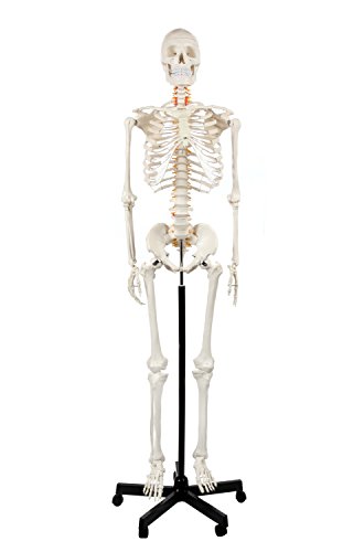
2. breesky Human Skeleton Model

The breesky skeleton stands taller than most at 70.8 inches, and its distinguishing feature is the included depiction of the nervous system overlaid on the skeletal frame. For students studying nerve root exits, spinal nerve pathways, or brachial plexus relationships, this visual overlay is a practical reference that keeps you from flipping back to a separate atlas.
The 206 articulated bones are cast from real human specimens, and reviewers consistently praise the anatomical accuracy of bony landmarks. The rolling stand with five casters makes repositioning effortless between study stations. Joints are detachable and movable, allowing you to isolate the forearm for supination/pronation drills or remove the skull for cranial nerve review.
Three posters are bundled with the model, covering skeletal anatomy, nerve distribution, and muscle attachment points. The PVC material cleans easily with a damp cloth. A few users noted the sacral attachment to the stand could be more robust, but the skeleton stays stable during normal use. This is a strong mid-premium option for visual learners who want nerve pathways mapped directly onto the bones.
Why it’s great
- Nervous system overlay on skeleton aids nerve pathway study
- Tallest model at 70.8 inches with smooth-rolling casters
- Accurate casting from real specimens
Good to know
- Sacral-to-stand connection could be stronger
- Not as many self-quizzing tools as the Axis Scientific
3. Giantex Life Size Human Skeleton

The Giantex skeleton earns its place in x-ray and imaging programs because of its accurate casting and fully articulating joints. Radiology and sonography students regularly rely on this model to correlate bone shadows on imaging studies with palpable landmarks. The detachable arms and legs allow focused study on individual joint complexes without the full skeleton in the way.
The movable jaw and removable skull cap give you access to the temporomandibular joint and cranial vault for deeper dissection-style review. Spinal nerves are molded into the vertebral column, which helps with nerve root identification. A stainless steel holder with casters makes it mobile, though assembly requires a hammer for the stand — plan for 20 minutes of setup.
The plastic construction is lightweight but durable, with a polished off-white finish. Some users reported a slightly warped pelvis and overly perfect denture-like teeth, but the overall anatomical proportions are accurate for study. It includes a plastic dust cover and anatomical chart. For the price, this skeleton punches above its weight for imaging-focused curricula.
Why it’s great
- Detachable limbs and movable jaw for focused study
- Molded spinal nerves aid nerve root review
- Lightweight and mobile with rolling stand
Good to know
- Stand assembly is challenging and requires a hammer
- Pelvis can be slightly asymmetrical
4. Evotech Disarticulated Human Skeleton

The Evotech disarticulated skeleton is the gold standard for osteology identification drills. Unlike fully assembled models that let you cheat by seeing adjacent bones, this set forces you to identify each bone in isolation — exactly what practical exams demand. The 67-inch set includes a full disarticulated skeleton plus an articulated left hand and foot for joint studies.
The skull is split into halves, allowing you to see both the external and internal cranial anatomy, including the sella turcica and the petrous ridge. Each bone includes accurate markings and textures that mimic real bone. The laminated poster labels all major bones, ribs, and vertebrae, serving as a quick reference during self-quizzing sessions.
One limitation: this model is not designed for assembly into a full standing skeleton. The ribs and thoracic cartilage lack the tools to lock together into a rigid cage. If your study method requires a mounted display, this may frustrate you. But for pure identification grinding — naming each bone, its markings, and its neighbors — this is the most effective tool in the list.
Why it’s great
- Disarticulated design forces active bone identification
- Split skull halve shows internal and external anatomy
- Articulated hand and foot included for joint study
Good to know
- Cannot be assembled into a mounted standing skeleton
- Some sharp mold seams require light sanding
5. Axis Scientific 34″ Life Size Spine Model

The Axis Scientific spine model is purpose-built for chiropractic, PT, and osteopathy students who need to understand vertebral articulation, spinal nerve exits, and the relationship between the lumbar curve and the pelvic tilt. At 34 inches and life-size, it includes the complete vertebral column with nerves, arteries, and a male pelvis.
The spine has moderate flexibility — enough to demonstrate normal curvatures and range of motion, but not so loose that it flops. The spinal nerves are color-coded and molded into the intervertebral foramina, making it easy to trace nerve roots from the cord to their peripheral pathways. The stand is lightweight but functional for desktop display.
Chiropractic students specifically praised this model for holding up to adjustment practice — it withstands moderate force without damaging the discs or vertebrae. The included full-color manual covers vertebral anatomy, nerve distribution, and common pathologies. For , this is the most cost-effective way to get a dedicated spine model that actually flexes and holds its shape.
Why it’s great
- Moderately flexible spine for adjustment practice
- Molded spinal nerves and arteries for pathway tracing
- Includes male pelvis for full lumbopelvic study
Good to know
- Stand is lightweight and tips easily
- Not as flexible as more expensive classroom models
6. EVOTECH SCIENTIFIC Human Body Model

The EVOTECH torso model is an 11-inch, 15-piece detachable mannequin that focuses on organ placement and spatial relationships. For students struggling to visualize how the liver sits beneath the diaphragm or the way the stomach connects to the esophagus, this hands-on model provides immediate clarity that no 2D diagram can match.
The set includes the trunk, brain, heart, lungs, stomach, liver, pancreas, spleen, intestines, and diaphragm. Each piece is hand-painted with detail and fits securely into its cavity — no collapsing organs. The oak-wood base provides a stable display platform, and the portable size fits into a standard backpack for transport to study groups or lab sessions.
The lungs are the most delicate component — some users reported they fall out during handling, which can be fixed with a small dab of museum wax. The pamphlet is basic and not particularly instructive. But as a visceral orientation tool for pre-clinical anatomy, this model fills a gap that skeleton-only study leaves wide open.
Why it’s great
- 15 removable organs teach spatial relationships
- Compact and portable for study group transport
- Stable oak-wood display base
Good to know
- Lungs may need museum wax to stay seated
- Included pamphlet is not very informative
7. Medarchitect Upgraded Life Size Skull Model

The Medarchitect skull solves the single most annoying problem with budget anatomical models: smudged annotations. Its laser-etched numbering is engraved into the PVC permanently — you can wipe it, clean it, and handle it constantly without a single number fading. For cranial foramina study, this is a critical advantage over hand-written models that become illegible after a few lab sessions.
The skull disassembles into three parts — calvaria, base of skull, and mandible — giving you access to the internal cranial floor. The mandible detaches easily, which is ideal for TMJ palpation practice. The 7.48 by 5.91-inch dimensions are true-to-life, and the non-toxic, tasteless PVC material has no off-gassing odor.
Some foramen features are slightly simplified compared to medical-grade casts, so you’ll want to cross-reference with an atlas for finer details like the cribriform plate foramina. The included instruction sheet lists features for memorization. For the price, this is the best entry-level skull for students who prioritize clean, permanent labeling over museum-quality casting.
Why it’s great
- Laser-etched numbers never smudge or fade
- 3-part disassembly reveals internal cranial floor
- Non-toxic PVC with no chemical smell
Good to know
- Some foramen details are simplified
- Mandible detaches very easily during handling
FAQ
Should I buy a full skeleton or a disarticulated model for medical school?
What is the most important feature to check on a skull model?
Can anatomy models substitute for cadaver dissection?
Final Thoughts: The Verdict
For most users, the anatomy models for medical students winner is the Axis Scientific Life Size Skeleton Model because its three study guides and 189 numbered stickers create a complete self-quizzing system that directly supports gross anatomy exam prep. If you want a spine model for chiropractic or PT study, grab the Axis Scientific 34″ Spine Model. And for focused bone-by-bone identification drills, nothing beats the Evotech Disarticulated Skeleton.